
קובץ:Modern 3T MRI.JPG

גודל התצוגה המקדימה הזאת: 800 × 600 פיקסלים. רזולוציות אחרות: 320 × 240 פיקסלים | 640 × 480 פיקסלים | 1,024 × 768 פיקסלים | 1,280 × 960 פיקסלים | 1,600 × 1,200 פיקסלים.
לקובץ המקורי (1,600 × 1,200 פיקסלים, גודל הקובץ: 204 ק"ב, סוג MIME: image/jpeg)
| זהו קובץ שמקורו במיזם ויקישיתוף. תיאורו בדף תיאור הקובץ המקורי (בעברית) מוצג למטה. |
| תיאור |
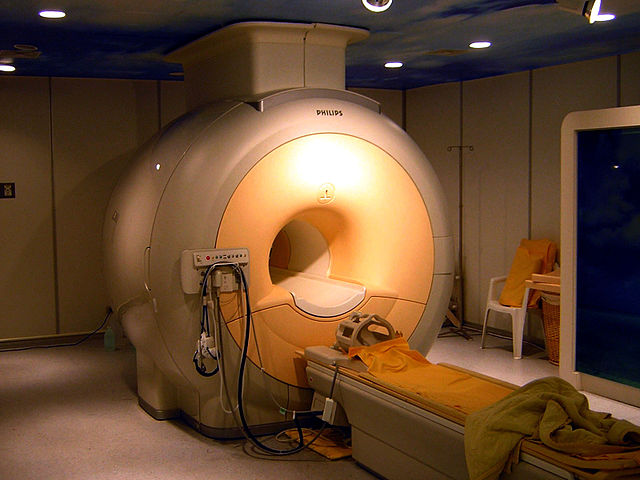
English: Modern high field clinical MRI scanner. (3T Achieva, the product of Philips at Best, the Netherlands.) |
||||||||
| תאריך יצירה | |||||||||
| מקור | Photographed by User:KasugaHuang on Mar 27, 2006 at Tri-Service General Hospital, Taiwan. | ||||||||
| יוצר | User:KasugaHuang | ||||||||
| אישורים והיתרים (שימוש חוזר בקובץ זה) |
KasugaHuang, בעל זכויות היוצרים על היצירה הזאת, מפרסם אותה בזאת תחת הרישיונות הבאים:
ייחוס: KasugaHuang
הנכם מוזמנים לבחור את הרישיון הרצוי בעיניכם. |

היסטוריית הקובץ
ניתן ללחוץ על תאריך/שעה כדי לראות את הקובץ כפי שנראה באותו זמן.
| תאריך/שעה | תמונה ממוזערת | ממדים | משתמש | הערה | |
|---|---|---|---|---|---|
| נוכחית | 18:11, 2 בדצמבר 2007 | | 1,200 × 1,600 (204 ק"ב) | Braegel | GIMP: Levels |
| 00:06, 4 באפריל 2006 |  | 1,200 × 1,600 (192 ק"ב) | KasugaHuang | Modern high field clinical MRI scanner. (3T Achieva, the product of Philips at Best, the Netherland.) Photographed by me on Mar 27, 2006 at Tri-Service General Hospital, Taiwan. |
שימוש בקובץ
הדף הבא משתמש בקובץ הזה:
שימוש גלובלי בקובץ
אתרי הוויקי השונים הבאים משתמשים בקובץ זה:
- שימוש באתר ar.wikipedia.org
- שימוש באתר ar.wikiversity.org
- שימוש באתר bg.wikipedia.org
- שימוש באתר bn.wikipedia.org
- שימוש באתר ca.wikipedia.org
- שימוש באתר ceb.wikipedia.org
- שימוש באתר ckb.wikipedia.org
- שימוש באתר da.wikipedia.org
- שימוש באתר de.wikipedia.org
- שימוש באתר de.wiktionary.org
- שימוש באתר el.wikipedia.org
- שימוש באתר en.wikipedia.org
- Engineering
- Helium
- Noble gas
- Niobium
- Superconducting magnet
- Portal:Engineering
- Instruments used in radiology
- User:The.Filsouf/Workspace
- User:Bci2/2D-FT NMR
- User:Bci2/Books/Wk1Book
- User:Garrondo/Sandbox/Interesting images
- Wikipedia:Recent additions/2009/January
- Physics of magnetic resonance imaging
- User:Bci2/Books/Biophysical Chemistry and Nuclear Medicine Vol 2
- User:Bci2/Books/Biophysical Chemistry Techniques and Nuclear Medicine Applications Vol 2
- User:Bci2/2D-FT NMRI and Spectroscopy
- User:Bci2/Books/NMRIS
- User:Daniel Mietchen/Talks/DigitalFossil Berlin 2012/Fossil MRI/MRI principle
- Monomelic amyotrophy